Why are facial fractures different?
The facial skeleton is not merely a framework that determines appearance. It protects the eyes, keeps the airway open, carries the teeth and distributes the forces of chewing. A facial fracture therefore affects function and appearance at the same time, and the aim of treatment is to restore both together.
Facial fractures most often follow traffic accidents, falls from a height, sports injuries, assaults and accidents at work. In recent years electric scooters and similar light vehicles have become a distinct cause of cranial and facial fractures, particularly in younger age groups.
Which bones are affected?
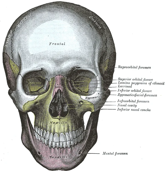
- The nasal bone. The most frequently fractured of the facial bones; it can leave a change in shape and nasal obstruction.
- The cheekbone and the eye socket. Fractures of the cheekbone flatten the face. The floor and inner wall of the orbit are thin; when they break, the eyeball can shift backwards or downwards.
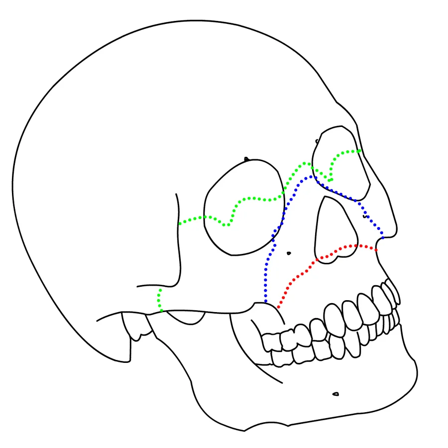
- The upper jaw. Classified according to the degree of displacement; it produces a disturbed bite and irregularity of the facial midline.
- The lower jaw and the jaw joint. These directly disturb the bite; restricted mouth opening and numbness of the lower lip may accompany them.
- The forehead and the frontal sinus. Their proximity to the brain and the optic nerves calls for particular care.
- Severe injuries involving the skull as well. Where several regions are fractured at once, treatment is conducted by a multidisciplinary team.
Which situations are emergencies?
The following findings call for going to hospital without delay:
- Reduced vision, double vision or restricted eye movement
- Difficulty breathing
- Clear, watery fluid coming from the nose or the ear
- Bleeding that cannot be stopped
- Clouded consciousness, severe headache or repeated vomiting
- Teeth that no longer meet, or inability to open the mouth
Signs
Depending on the site of the fracture, the following are seen: swelling and bruising of the face, bleeding around the eye, flattening over the cheekbone, numbness of the cheek and upper lip, a change in the bite, restricted mouth opening, and a change in the shape of the nose with obstruction.
Swelling can mask the underlying deformity in the first days. Reassessment once the swelling has settled is therefore important for building the treatment plan correctly.
Diagnosis
On examination, facial symmetry, eye movements, skin sensation, the bite and mouth opening are assessed systematically.
For imaging, fine-slice computed tomography with three-dimensional reconstruction is essential; it shows the fracture lines, the number of fragments and the extent of displacement. In fractures involving the orbit an ophthalmological assessment, and in jaw fractures a detailed examination of the bite, determine the treatment decision.
Treatment
Not every fracture requires surgery. Fractures that are undisplaced and do not disturb the bite or eye function can heal with close follow-up, a soft diet and pain control.
Surgery is decided upon when there is displacement of the fragments, disturbance of the bite, ocular findings, difficulty breathing or marked asymmetry.
- Timing. Surgery is usually planned once the swelling has settled but before bony union begins. Conditions threatening the eye or the airway fall outside this sequence and are brought forward.
- Incisions. Intraoral routes and natural creases (the eyebrow, the inner eyelid, the hairline) are used to keep scars away from visible areas.
- Fixation. The fragments are returned to their anatomical position and secured with titanium plates and screws compatible with magnetic resonance imaging.
- Orbital repair. The thin walls that have broken are reconstructed with a suitable implant or bone graft; the aim is to preserve the position and movement of the eyeball.
- Jaw fractures. The correct bite serves as the reference; elastics or an arch bar may be used for a period where required.
- Approach in children. Because growth continues, treatment is more conservative; resorbable plates are used in suitable cases and facial development is followed over a long period.
Recovery
Swelling decreases markedly within the first week or two. During this period a soft or liquid diet is followed and particular attention is paid to oral hygiene.
In fractures of the upper jaw and the orbit, simple precautions matter — not blowing the nose forcefully, keeping the mouth open when sneezing — otherwise air can track into the tissues. Bone healing is usually largely complete within six weeks, although changes in sensation and the last of the swelling may take longer to resolve.
Late corrections
Where a fracture has united but healed in the wrong position, facial asymmetry, a sunken eyeball, a disturbed bite or a deformity of the nose may remain.
These can be corrected by secondary reconstruction: planned re-cutting of the united bone, bone grafting, reconstruction of the orbit and, where needed, orthognathic surgery. Three-dimensional planning markedly improves accuracy in late corrections.
Assessment
Two things determine the outcome in facial fractures: mapping the fracture correctly and operating at the right time. Two fractures that look alike may carry different functional risks.
If you have a change in facial shape, numbness or a problem with your bite after an injury — whether the fracture is recent or old — you are welcome to book an appointment for assessment.