What is craniosynostosis?
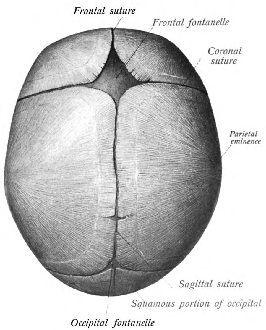
An infant’s skull is not a single piece of bone. So that it can expand during the first years, when the brain grows fastest, the skull bones are joined by flexible seams called sutures. These sutures normally close gradually, over years, as brain growth is completed.
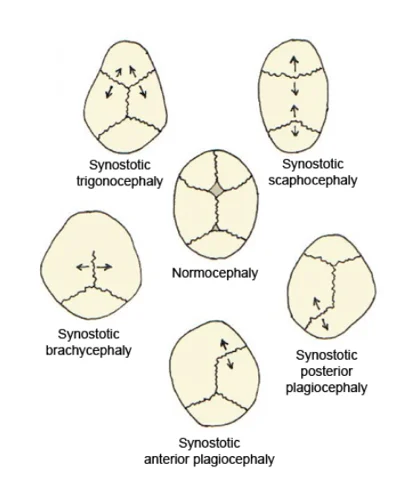
In craniosynostosis, one or more sutures close far earlier than they should — usually before birth or within the first months of life. The skull can no longer grow across the fused suture, so growth is redirected to the sutures that remain open. The result is a head that develops in a characteristically asymmetric way.
This is not only a question of appearance. As the number of fused sutures increases, the volume of the skull may no longer meet the needs of the growing brain and intracranial pressure can rise.
What are the signs?
Craniosynostosis usually draws attention in the first months through a change in head shape. The findings parents and physicians look for include:
- Elongation in one direction, flattening at the sides, or clear asymmetry of the forehead or temples
- A firm ridge that can be felt along the fused suture line
- A fontanelle that closes earlier than expected or feels unusually tense
- Head circumference that fails to follow the expected growth curve
- Differences in the shape of the eye sockets and the midface
- Irritability, feeding difficulty, vomiting or loss of developmental milestones, which may point to raised intracranial pressure
Not every irregular head shape is craniosynostosis. Positional head shape deformities, which develop when a baby consistently lies on the same side, are far more common and do not require surgery. Distinguishing between the two is the first and most important step in treatment.
How is the diagnosis made?
Assessment begins with a detailed physical examination: mapping the head shape, palpating the suture lines, plotting head circumference against the growth curve, and an eye examination.
Three-dimensional computed tomography (CT) is generally used to confirm the diagnosis and plan surgery; it shows precisely which sutures have fused and the three-dimensional geometry of the skull. Where helpful, skull modelling supports preoperative planning.
Craniosynostosis sometimes occurs on its own and sometimes as part of a syndrome. If there are additional findings involving the hands, feet, face, heart or hearing, genetic assessment and the opinion of the relevant specialties are requested.
Surgical treatment
Surgery has two aims: to give the growing brain adequate volume, and to make the shape of the head and face as symmetric as possible. The technique is chosen according to which suture has fused, the age of the baby, the state of intracranial pressure and any associated anomalies.
- Endoscope-assisted procedures. In suitable cases diagnosed early, the fused suture is released through small incisions. Operating time and blood loss are lower; treatment is usually supported by a moulding helmet.
- Open cranial vault remodelling. The affected part of the skull is removed, reshaped and replaced. This is preferred when several sutures have fused and more extensive correction is required.
- Distraction (gradual bone lengthening). An implanted device moves the bone segments apart in small, controlled daily steps, increasing skull volume progressively.
Surgery is performed with a plastic surgeon and a neurosurgeon working together, in a setting with an anaesthetic team experienced in infant anaesthesia and blood management. Because suture fusion progresses from the skull base upwards, planning covers not only the cranial vault but the orbits and the midface as well.
Why does timing matter?
Craniosynostosis surgery is usually planned during infancy, within the first year of life. At this stage the bone is easier to reshape and the capacity for healing is at its greatest.
If there are signs of raised intracranial pressure, swelling of the optic disc (papilloedema), or progressive loss of vision or hearing, assessment and treatment become urgent. Timing is nevertheless not identical for every baby; early diagnosis matters because it widens the options.
Recovery
The first 24–48 hours are usually spent under close monitoring in intensive care. The expected swelling of the face and around the eyes settles within a few days. Babies are generally discharged within a few days, and feeding and sleep patterns return to normal soon afterwards.
Follow-up does not end with a single operation. Because the skull and face continue to grow, the child is reviewed at regular intervals for head shape, intracranial pressure, vision, hearing and speech development. This follow-up makes it possible to plan any further correction at the right time.
Assessment
The degree of craniosynostosis, which sutures have fused and the risks that follow differ in every baby. The right approach is decided after examination and imaging, through planning that includes the family.
If you have any concern about your child’s head shape, you are welcome to book an appointment for assessment.